.png)
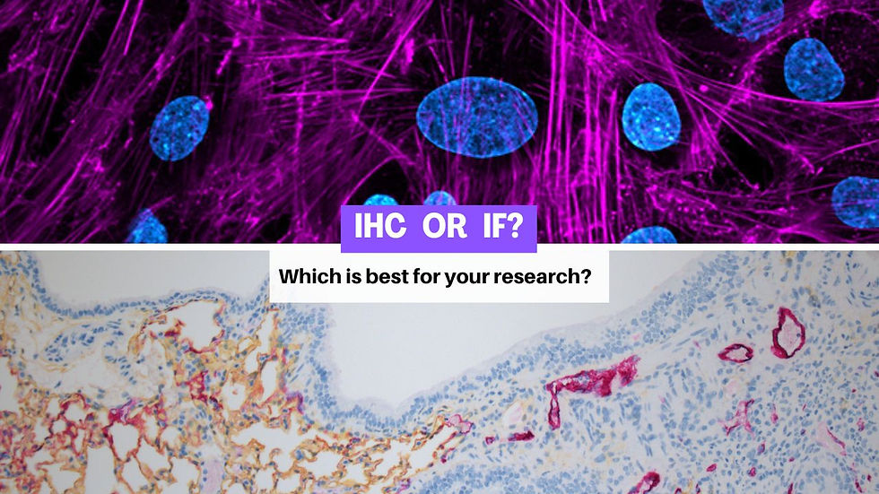
IHC vs IF: Which Is Better for Your Tissue Research?
- Eghosa Arovo

- Feb 16
- 4 min read
IHC or IF? Here’s how to choose the right technique for your tissue research.
Introduction
When planning a tissue-based research project, one of the most common questions researchers ask is:Should I use immunohistochemistry (IHC) or immunofluorescence (IF)?
Both techniques use antibodies to detect proteins in tissue, and both are powerful tools in cancer and tissue research. However, they differ significantly in practicality, robustness, interpretability, and long-term usability.
In this blog, we compare IHC vs IF and explain why—for most research applications—chromogenic IHC remains the more reliable, scalable, and cost-effective choice.
Quick recap: What’s the difference?
Immunohistochemistry (IHC)Uses enzyme-linked antibodies and chromogens (e.g. brown DAB, red, purple, teal, yellow) to produce coloured precipitates at antigen sites. Slides are viewed using a standard brightfield microscope.
Immunofluorescence (IF)Uses fluorescently labelled antibodies (fluorophores) that emit light under specific wavelengths and must be visualised using a fluorescence microscope or scanner.
Both detect proteins accurately—but the way the signal behaves over time, and how easily it integrates into real research workflows, is where the key differences appear.
Why IHC is usually the better choice for research
1. Permanent, archive-ready slides
One of the biggest practical advantages of chromogenic IHC is signal stability.
DAB and other chromogens form insoluble, permanent precipitates
Slides can be stored for years without signal loss
Data can be re-reviewed, rescanned, or published long after staining
By contrast, IF signals fade over time (photobleaching), especially with repeated imaging or long-term storage. This makes IF less suitable for retrospective studies, biobanking projects, and long-term datasets.(Bancroft & Gamble, 2020)
2. Easier interpretation and validation
IHC slides are:
Viewed in brightfield
Interpretable by anyone trained in histology
Directly comparable to decades of published literature
Most antibody datasheets, validation studies, and scoring systems are built around IHC-DAB. That means your results are easier to benchmark, validate, and publish.
IF, while powerful, often requires:
Careful spectral compensation
Autofluorescence correction
Specialised imaging and software
This adds complexity and variability—especially across labs and projects.
3. Lower technical and financial barriers
Chromogenic IHC requires:
Standard microscopes
Simple image capture
Lower-cost reagents and consumables
IF requires:
Fluorescence microscopes or scanners
Expensive fluorophores
Ongoing calibration and maintenance
Dark storage and careful handling to prevent signal loss
For most research teams, especially in academic and translational settings, IHC delivers far better cost-to-value.
4. Excellent compatibility with multiplexing
Many researchers assume that IF is required for multiplexing. In reality, chromogenic multiplex IHC is now highly advanced.
Using different chromogens—such as:
Red, Purple, Teal, Blue, Green, Yellow, DAB, and Silver
You can visualise multiple antigens on the same tissue section with excellent contrast.
Even better, chromogenic multiplex enables the “1 + 1 = 3” effect:when two colours co-localise, they produce a third colour—making double-positive cells visually obvious without complex software.
This allows robust multiplexing while retaining all the benefits of brightfield histology.(Maiques et al., 2022)
5. Ideal for digital pathology and AI analysis
Chromogenic IHC is highly compatible with:
Whole-slide scanning
Quantitative image analysis
AI-based segmentation
Long-term dataset generation
Clear colour separation and stable signal make chromogenic slides easier to analyse reproducibly than fluorescence images, which can vary with scanner settings, exposure time, and signal decay.
When does IF make sense?
To be balanced, IF does have real strengths.
You may prefer IF if you:
Need extremely high sensitivity for very low-abundance proteins
Require ultra-high multiplexing (6–20+ markers)
Are working in subcellular localisation or signalling studies
Already have a fully optimised fluorescence imaging pipeline
IF is also valuable in exploratory or mechanistic studies—but it is rarely the most practical option for routine, scalable, or translational histology research.
Why most researchers still choose IHC
For the majority of cancer, immunology, and tissue-based projects, IHC offers the best balance of:
Robustness
Interpretability
Reproducibility
Cost efficiency
Archival stability
That’s why it remains the global workhorse of histology—even as multiplex and spatial biology workflows evolve.
LabNexus: Your IHC specialist partner
At LabNexus, we are purpose-built to support high-quality chromogenic IHC for research.
We offer:
Single-plex IHC using classic DAB
Single-plex IHC using bright chromogens(Red, Purple, Teal, Blue, Green, Yellow, Silver)
Multiplex chromogenic IHC panels
Tissue processing, embedding, and sectioning
Slide scanning for digital analysis
All IHC staining is performed using our state-of-the-art Ventana Benchmark platforms, delivering:
High reproducibility
Excellent signal-to-noise ratios
Clean background
Consistent turnaround times
This means you get the power of multiplex and colour—without the complexity and instability of fluorescence.
Please note: LabNexus provides histology services for research purposes only. We do not process diagnostic samples.
Conclusion
Both IHC and IF are valuable tools—but for most tissue research projects, chromogenic IHC is the smarter default choice.
It offers:
Permanent, archive-ready results
Easier interpretation and validation
Lower technical and financial burden
Robust multiplexing capability
Seamless integration with digital pathology
Whether you need classic DAB IHC, bright coloured stains, or multiplex panels, LabNexus can support your research with high-standard histology services and fast turnaround times.
References
Bancroft, J.D., & Gamble, M. (2020). Theory and Practice of Histological Techniques (8th ed.). Elsevier.
Abcam. Immunohistochemistry (IHC) staining guide.https://www.abcam.com/en-us/knowledge-center/immunohistochemistry/ihc-staining
Abcam. Immunofluorescence (IF) staining overview.https://www.abcam.com/en-us/knowledge-center/immunofluorescence/if-staining
Maiques, O. et al. (2022). Multiplex chromogenic immunohistochemistry to stain and analyse two markers in paraffin tissue sections. MethodsX, 9, 101788.
Ramos-Vara, J.A. (2005). Technical aspects of immunohistochemistry. Veterinary Pathology, 42(4), 405–426.


Comments