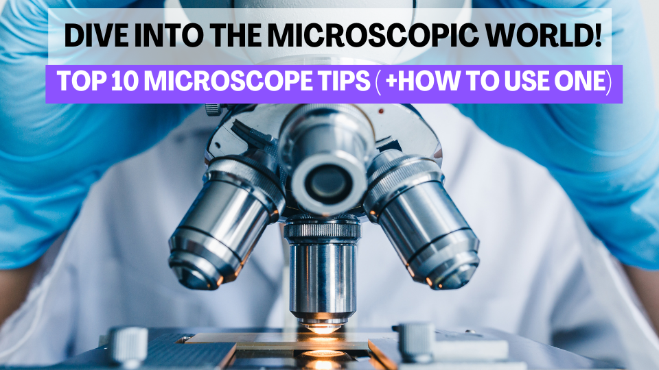
Top 10 Tips & Tricks for Better Imaging with Microscopes
- Eghosa Arovo

- Mar 10
- 3 min read
Want to master microscopy? Learn how to set up, focus, and capture the best images with our Top 10 Microscopy Tips & Tricks! 📖👇 #Microscopy #Histology #LabNexus
Microscopy is a fundamental tool in histology and biological research, allowing scientists to examine cells, tissues, and microorganisms at a microscopic level. Whether you're a beginner or an experienced researcher, understanding how to use a microscope correctly is essential for producing high-quality images and making accurate observations.

Getting Started: How to Use a Microscope
Before diving into advanced microscopy tips, it's important to understand the basic steps of using a microscope properly.
1. Setting Up the Microscope
Place the microscope on a stable, flat surface to avoid vibrations.
Ensure the light source is working and adjust the brightness before inserting the slide.
Make sure the stage is clean and the lenses are free of dust or fingerprints.
2. Placing the Slide
Hold the slide by the edges to avoid fingerprints on the specimen area.
Place the slide on the stage and secure it using the stage clips.
Use the mechanical stage knobs to center the specimen under the objective lens.
3. Adjusting the Objective Lenses
Start with the lowest magnification objective (4x or 10x).
Use the coarse focus knob to bring the specimen into rough focus.
Switch to higher magnification objectives only after the specimen is centered and in focus.
Use the fine focus knob for sharp and clear imaging.
4. Adjusting the Light and Contrast
Use the iris diaphragm to control the amount of light passing through the specimen.
Properly align the condenser to enhance image contrast.
Apply Köhler illumination for uniform lighting.
5. Focusing Techniques
When using high magnification (40x or 100x oil immersion lenses), always fine-tune the focus slowly to avoid damaging the slide.
Keep both eyes open to reduce eye strain while adjusting the focus.
The Top 10 Microscopy Tips & Tricks for Better Imaging
1. Keep Your Microscope Clean
A dirty microscope can lead to blurry images and inaccurate results. Regularly clean the objective lenses, eyepieces, and stage with lens paper and ethanol to prevent dust and oil buildup.
2. Adjust the Lighting Properly
Use Köhler illumination to evenly distribute light and reduce glare. Adjust the brightness settings to avoid overexposure.
3. Use the Right Objective Lens
Different samples require different magnifications. Start with low magnification (4x or 10x) to locate your sample, then switch to higher magnifications for detailed analysis.
4. Focus Carefully
Use coarse focus at low magnifications.
Switch to fine focus for higher magnifications.
Adjust focus slowly to prevent damage to the slide and objective lenses.
5. Apply the Correct Contrast Techniques
Contrast helps differentiate structures within the sample. Depending on your specimen, consider using:
Phase contrast for transparent specimens.
DIC (Differential Interference Contrast) for more detailed 3D-like imaging.
Staining methods like H&E or IHC for enhanced visualization.
6. Minimize Air Bubbles in Slide Preparation
Bubbles can distort your view and affect image quality. When mounting a sample, slowly lower the cover slip at an angle to reduce trapped air.
7. Calibrate the Microscope Regularly
Ensure accuracy by calibrating the stage micrometer and adjusting the focus alignment. This is especially important for measuring structures in histology.
8. Store Microscope Slides Properly
Prevent sample degradation by storing slides in a dry, dust-free environment. For long-term preservation, keep them in labeled slide boxes at room temperature.
9. Take Advantage of Digital Microscopy
If available, use slide scanning or digital imaging software to capture high-resolution images. Digital storage makes it easier to analyze, share, and compare results.
10. Handle the Microscope with Care
Microscopes are delicate instruments. Always handle the objective lenses with care, avoid unnecessary pressure on knobs, and store the microscope covered when not in use.
Conclusion
Mastering microscopy is essential for producing clear, detailed images that support research and diagnostics. By following these basic principles and advanced tips, you can improve your microscopy skills and ensure accurate observations.
At LabNexus, we provide expert histology services, including slide preparation, staining, and imaging support. If you need high-quality histological analysis for your research, contact us today!
.png)


Comments