.png)
Beyond Brown: Using Colourful IHC Chromogens to Power Your Histology Research
- Eghosa Arovo

- Feb 2
- 5 min read
Move past brown DAB: discover how colourful IHC chromogens can unlock more insight from your tissue sections.

Quick recap: What IHC does for your tissue research
In last week’s blog, we looked at immunohistochemistry (IHC) as a way to detect specific proteins in tissue using antibodies. By binding a primary antibody to an antigen, then visualising it with an enzyme–chromogen system, IHC lets you see where a protein is expressed in the tissue and how much is present.
Most research labs (and many histology services in the UK) still use a single brown chromogen, DAB (3,3′-diaminobenzidine), to reveal this signal. DAB is reliable, robust and easy to interpret, but it’s not the only option. Once you start thinking in colour, you can extract much more bandwidth from each slide.
From DAB to colour: why use different chromogens?
In chromogenic IHC, enzymes such as HRP or alkaline phosphatase convert a soluble substrate into a coloured, insoluble precipitate at the antigen site. DAB gives a brown signal with HRP and has become the “default” because it’s stable in organic solvents and works beautifully with a haematoxylin counterstain.
However, modern chromogen ranges now include bright, high-contrast colours—ideal for:
Highlighting subtle structures that get lost in brown-on-pink H&E backgrounds
Double- or multi-plex IHC, where you need to distinguish several markers in the same section
Digital image analysis, where clear colour separation improves segmentation
At LabNexus, alongside classic DAB we offer a bright palette of chromogens for research IHC:
Red, Purple, Green, Yellow, Silver, Teal
These can be used individually (single-plex IHC) or in carefully chosen combinations for dual or multiplex chromogenic IHC.

Colour mixing and co-localisation: when 1 + 1 = 3
One of the big advantages of coloured chromogens is that co-localisation produces a third colour. When two antigens are present in the same cell or structure, their chromogens overlay optically and create a mixed colour, making double-positive populations easy to spot. This principle is widely used in multiplex chromogenic IHC design.
Example: proliferating tumour cells
Imagine you’re studying a solid tumour and you want to see:
Pan-cytokeratin – to mark epithelial tumour cells
Ki-67 – to mark proliferating nuclei
You could design a dual-colour chromogenic IHC like this:
Ki-67 detected with a red chromogen
Pan-cytokeratin detected with a yellow chromogen
On your section you would see:
Cytokeratin-positive but Ki-67-negative tumour cells in yellow
Ki-67-positive but cytokeratin-negative cells (e.g. some stromal cells) in red
Double-positive, actively proliferating tumour cells in orange (red + yellow)
That third colour (orange) instantly flags the cells you care about most—no extra serial sections, no guessing which nuclei belong to which cells.
The same logic can be applied to many biologically relevant pairs: for example, CD3 with FOXP3 for regulatory T cells, or CD68 with PD-L1 in macrophage-rich tumour microenvironments.
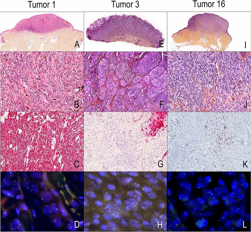
Case example: Dual-colour IHC for stromal activation and tumour proliferation

The image above shows a dual-colour immunohistochemistry (IHC) stain using two widely used cancer research markers: α-smooth muscle actin (α-SMA) and Ki-67.
Take α-smooth muscle actin (α-SMA), a classic marker of:
Vascular smooth muscle cells
Pericytes
Myofibroblasts in fibrotic or tumour stroma
If α-SMA is stained with brown DAB, it can sometimes blend into collagen-rich stroma or pigmented areas, especially once counterstained with haematoxylin. Using a red chromogen instead:
The α-SMA-positive cells stand out clearly as red cytoplasm against a blue–purple nuclear counterstain
Blood vessels and myofibroblast-rich regions become easier to segment for image analysis
In multiplex panels, red α-SMA can be visually separated from, for example, a teal immune marker or yellow epithelial marker
At LabNexus we routinely run α-SMA with red chromogen for research projects that focus on stromal activation, fibrosis, or angiogenesis, particularly in cancer and tissue-engineering models.
Why is DAB still the workhorse chromogen?
With all these colours available, it’s a fair question: why do most research histology labs still default to DAB?

There are several reasons:
Stability and archivingDAB precipitate is highly stable, survives dehydration in alcohol and clearing in xylene, and can be mounted in standard permanent media. Slides remain interpretable for years, which is ideal for long-term studies and retrospective analysis.
Compatibility and traditionMost published IHC protocols, antibodies, and validation studies assume DAB. Using it makes your data easier to compare with existing literature, and many analysis tools are tuned to brown-on-blue images.
Sensitivity and contrastDAB with HRP is very sensitive and provides strong nuclear or cytoplasmic contrast with a simple haematoxylin counterstain. It works well for both low- and high-expressing antigens.
Practical considerationsSome coloured chromogens (like AEC or Fast Red) are alcohol-soluble and require aqueous mounting media; they can also be more sensitive to light fading. This is fine for many research applications but less convenient for routine archiving or shipping slides.
For these reasons, many researchers still choose DAB for single-plex IHC and reserve bright chromogens for special cases or multiplex panels.
Putting it together: designing smarter IHC for your project
When you plan your next experiment, think of IHC not just as “brown vs negative”, but as a flexible colour toolkit:
Use DAB when you need robust, archive-ready slides and straightforward single-marker assessment.
Use bright chromogens when:
You need better contrast on a tricky background
You’re running double or multiplex IHC
You plan downstream digital or AI-based analysis that benefits from strong colour separation
At LabNexus, we can help you design IHC panels that make the most of your tissue sections—whether that’s single-colour DAB, dual-colour chromogenic IHC, or more complex multiplex strategies combined with TMAs (tissue microarrays) to increase throughput.
Conclusion
Immunohistochemistry is already a powerful technique for mapping proteins in tissue. By moving beyond a single brown chromogen to a palette of bright colours, you can:
Detect multiple antigens in the same section
Visualise co-localisation as intuitive third colours
Improve contrast for challenging markers like α-SMA in dense stroma
Prepare your slides for quantitative, digital analysis
Whether you want classic DAB IHC or to explore our range of bright chromogens—red, purple, green, yellow, silver, teal—LabNexus offers a full suite of research-only histology services, including IHC panel design, staining, slide scanning and TMA-based multiplex workflows.
If you’re planning a project and would like advice on which chromogens or multiplex strategy suits your research, our team is happy to help.
Book a free consultation with LabNexus.
References
Images obtained from LabNexus and partner's in-lab stained slides.
Abcam. Immunohistochemistry (IHC) staining guide.
Agilent Technologies. Immunohistochemical Staining Methods.
R&D Systems. Chromogenic IHC staining protocol for paraffin-embedded tissue.
Leica Biosystems. Tips & Tricks to Multiplexing: How to Choose Chromogen Colors for Multiplex.
Abcam. Multiplex immunohistochemistry (mIHC): technical deep dive.
Maiques, O. et al. Multiplex chromogenic immunohistochemistry to stain and analyse two markers in paraffin tissue sections. MethodsX (2022).
Sigma-Aldrich. Actin, α-Smooth Muscle, Immunohistology Kit – staining characteristics.


Comments