.png)
Immunohistochemistry (IHC): What It Is and Why It Matters for Tissue Research
- Eghosa Arovo

- Jan 30
- 4 min read
New to IHC? Here’s what it is, why it matters, and how LabNexus can support your research.

Introduction
Immunohistochemistry (IHC) is one of the most powerful and widely used techniques in modern histology and tissue research. From cancer biology to immunology and developmental studies, IHC allows researchers to visualise where specific proteins are expressed within tissue, and how those proteins relate to structure, disease, and cell behaviour.
This blog marks the start of a new IHC-focused series at LabNexus. Over the coming weeks, we’ll explore how IHC works, how to design staining panels, how to use colour and multiplexing, and how to extract the most meaningful data from your tissue sections.
We begin with the fundamentals: what IHC is, why it is so useful in research, and how LabNexus can support your IHC workflows end to end.
What is immunohistochemistry (IHC)?
Immunohistochemistry (IHC) is a technique that uses antibodies to detect specific proteins (antigens) in tissue sections.
In simple terms, it works like this:
A primary antibody binds to a protein of interest in the tissue.
A secondary antibody binds to the primary antibody.
An enzyme linked to the secondary antibody converts a substrate into a visible coloured precipitate (such as brown DAB or a coloured chromogen).
The result is a stained tissue section showing exactly where the target protein is located.

This allows researchers to see protein expression in spatial context, rather than as an average signal from homogenised tissue.
Why is IHC so useful in research?
IHC remains the global workhorse of histology research for several key reasons.
1. It preserves tissue architecture
Unlike Western blotting or PCR, IHC shows where proteins are located within the tissue. You can distinguish expression in:
Tumour cells vs stromal cells
Immune cells vs epithelium
Nuclei vs cytoplasm vs membrane
This spatial context is critical for cancer research, immune profiling, and tissue biology.
2. It is highly interpretable
Chromogenic IHC produces coloured signals that are:
Easy to visualise in brightfield microscopy
Compatible with decades of published literature
Suitable for both qualitative and quantitative analysis
Markers such as Ki-67, p53, CD3, CD8, and PD-L1 are routinely assessed using IHC because the results are intuitive and biologically meaningful.
3. It scales from simple to advanced
IHC is extremely flexible:
Single-plex IHC for one marker (e.g. Ki-67 or p16)
Coloured IHC for better contrast
Multiplex chromogenic IHC for detecting multiple markers on one slide
Integration with TMAs for cohort studies
This makes IHC suitable for both small pilot projects and large translational studies.
Why antibody optimisation matters
One of the most overlooked steps in immunohistochemistry (IHC) is antibody optimisation. Not all antibodies behave well in fixed tissue, and even those that are “validated” still require careful tuning to work properly under specific experimental conditions. Key factors such as antigen retrieval, antibody dilution, incubation time, detection chemistry, and background suppression all need to be adjusted to achieve a clean, specific, and interpretable signal.
Without proper optimisation, IHC results can be weak or falsely negative, overstained or nonspecific, and inconsistent between runs. This makes data difficult to interpret and can compromise the reliability of an entire study. For this reason, antibody optimisation is a critical step that should always be completed before staining valuable experimental samples.
How LabNexus supports IHC antibody optimisation
At LabNexus, we provide a full IHC optimisation service for research antibodies.
You can either:
Provide your own control tissue, or
Ask us to assess whether we already have appropriate control tissue in-house.
We then run a structured optimisation workflow to determine:
The best antigen retrieval method
The optimal antibody concentration
The most suitable detection system
The cleanest signal-to-noise conditions
Once your antibody is optimised, the protocol can be locked down and reused reliably across future experiments.
End-to-end histology and IHC at LabNexus
LabNexus offers a complete research histology service — from tissue arrival to stained slides.
We can support your project with:
Tissue processing
Processing of human and animal research tissues
FFPE workflows
Research-only samples (non-diagnostic)
Paraffin embedding
Standard embedding
Custom orientations
Embedding of blank recipient blocks (for TMA workflows)
Sectioning
Flexible section thickness
Any number of sections per block
Full-section or TMA sectioning
IHC staining
Single-plex IHC with classic DAB
Single-plex IHC with coloured chromogens
Multiplex chromogenic IHC panels
Antibody optimisation and protocol development
Why start your IHC journey with LabNexus?
At LabNexus, we specialise in research-only histology and IHC services for cancer and tissue researchers across London and the UK.
We offer:
Antibody optimisation support
Control tissue sourcing or evaluation
End-to-end histology workflows
Automated, high-quality IHC staining
Single-plex and multiplex chromogenic IHC
Fast turnaround times
Affordable pricing
Please note: LabNexus provides histology services for research purposes only. We do not process diagnostic samples.
Conclusion
Immunohistochemistry is one of the most powerful tools in tissue research — allowing you to visualise proteins in their true biological and spatial context.
When combined with proper antibody optimisation, automated staining, and high-quality histology workflows, IHC becomes a robust, scalable, and highly informative research technique.
This blog marks the beginning of our IHC education series at LabNexus. In the coming posts, we’ll explore coloured IHC, multiplex panels, antibody optimisation strategies, and advanced applications for cancer and tissue research.
If you’re planning an IHC project and need help optimising antibodies or processing and staining your tissue, LabNexus is here to support your research.
References
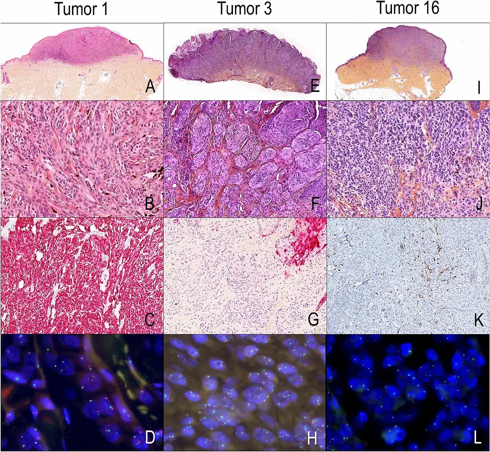
Slide section images were from samples processed, embeded, cut and stained by LabNexus and partner laboratories.
How IHC Works [Image] by OncoDaily: https://oncodaily.com/oncolibrary/immunohistochemistry
Bancroft, J.D., & Gamble, M. (2020). Theory and Practice of Histological Techniques (8th ed.). Elsevier.
Ramos-Vara, J.A., & Miller, M.A. (2014). When tissue antigens and antibodies get along: Revisiting technical aspects of immunohistochemistry. Veterinary Pathology, 51(1), 42–87.
Abcam. Immunohistochemistry (IHC) staining guide.https://www.abcam.com/en-us/knowledge-center/immunohistochemistry/ihc-staining
Taylor, C.R., & Shi, S.R. (2013). Antigen retrieval in immunohistochemistry: Past, present, and future. Journal of Histochemistry & Cytochemistry, 61(1), 12–32.


Comments