.png)
Why Histology is Essential for Tissue Research
- Eghosa Arovo

- Jan 28
- 3 min read
Tissue research? Here's why histology is your most powerful tool for understanding what’s really happening at the cellular level.

Introduction
Behind every tissue-based research breakthrough—whether in cancer biology, regenerative medicine, or developmental studies—there’s one common tool: histology.
Histology, the microscopic study of tissues and cells, remains one of the most powerful, cost-effective, and widely used techniques in biological and medical research. It provides insights that imaging, molecular, and in vitro methods often miss, and when combined with other tools, it becomes a pillar of translational science.
In this blog, we break down why histology is so beneficial for tissue research, how researchers can best take advantage of it, and how LabNexus can support you with high-quality, research-only histology services in London and across the UK.
What Makes Histology So Important in Tissue Research?
1. Visualising Tissue Structure and Cellular Architecture
Histology allows researchers to examine:
The overall organisation of tissue
The types of cells present
How cells interact with their environment
Whether tissue is normal, inflamed, fibrotic, or neoplastic
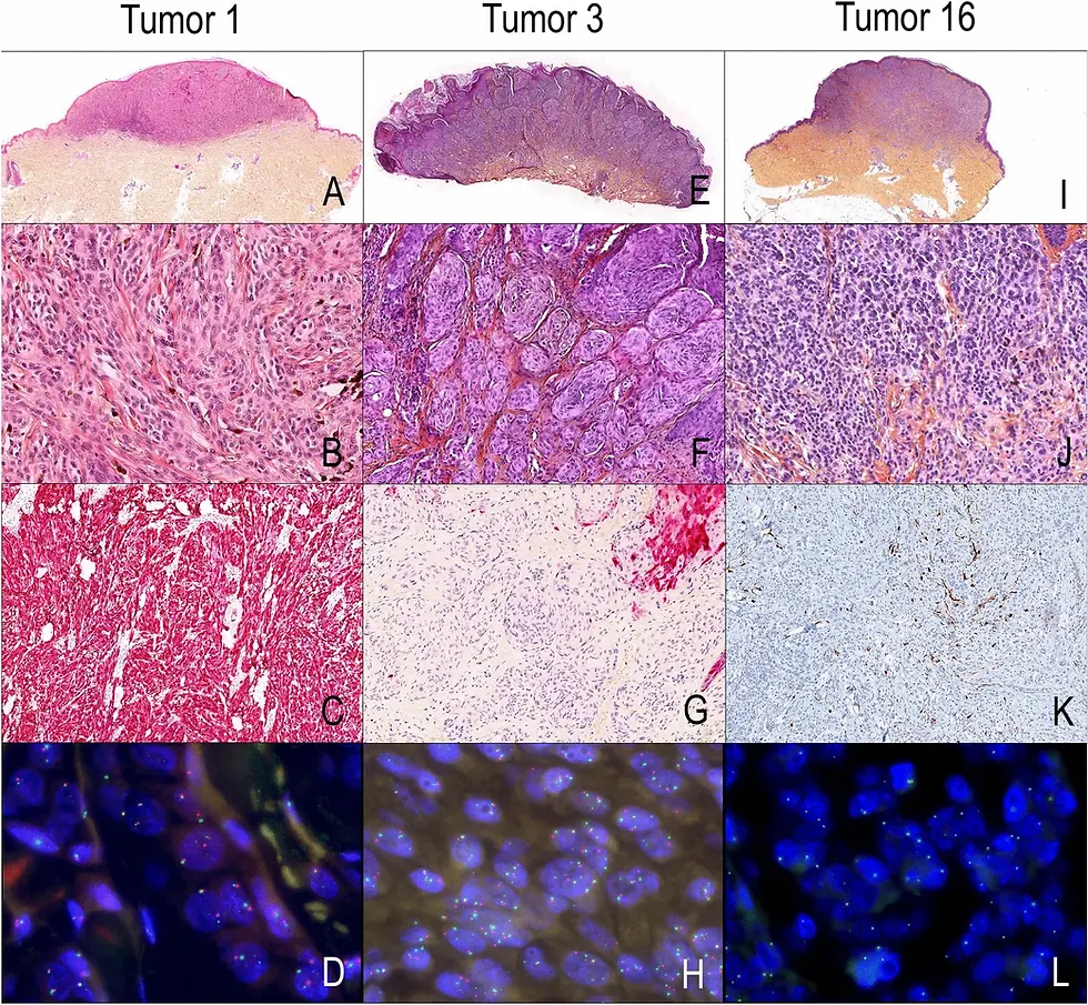
These details are critical in research areas like tumour progression, wound healing, organ development, and fibrosis studies. A standard Hematoxylin & Eosin (H&E) stain can reveal architecture at a glance, while special stains and IHC provide deeper insights into protein expression and structure.
2. Confirming Experimental Outcomes
Whether you're testing a new drug, implant, or gene therapy, histology helps you:
Confirm whether your intervention worked
Check for off-target effects or toxicity
Measure tissue regeneration or degeneration
Quantify inflammatory responses or fibrosis
Without histology, it’s hard to prove that a treatment had a true structural or cellular impact. It provides visual proof, often required for publications or regulatory reporting.
3. Understanding Disease Mechanisms
In cancer and pathology-related studies, histology offers vital clues:
Tumour grading and staging through cell differentiation and mitotic index
Invasion patterns, necrosis, and angiogenesis
Stromal and immune cell interactions, especially in the tumour microenvironment
Histology bridges macroscopic observations (what we can see with the eye) with molecular findings, making it essential in understanding disease progression and response.
4. Supporting Quantitative and Digital Analysis
Modern histology goes beyond observation. With digital slide scanning, researchers can:
Quantify collagen content, vessel density, or cell counts
Use AI-powered tools for pattern recognition
Store and share high-resolution slides for collaboration
Digital histology makes tissue data more reproducible and scalable, especially when combined with image analysis software.
What Types of Histology Are Common in Tissue Research?
LabNexus supports a wide range of research projects, and the most common histological techniques include:
H&E Staining – the universal starting point for tissue assessment
Special Stains – such as Sirius Red (for collagen), Perls’ (for iron), DPAS (for carbohydrates), Masson’s Trichrome (for fibrosis), and EVG (for elastic fibres)
Immunohistochemistry (IHC) – to identify and localise proteins, immune markers, or disease-related targets
Cryosectioning – for fast analysis of fresh/frozen tissues and delicate epitopes
Whether you work with human or mouse tissue, these techniques can be tailored to your study goals.
Why Work With a Research-Only Histology Partner?
At LabNexus, we specialise in histology services for research purposes only. That means:
No diagnostics
No long waiting lists
Full flexibility based on your research needs
We cater to PhD students, postdocs, PIs, and biotech researchers who need expert histology support but don’t have access to their own lab or technicians.
With LabNexus, you get:
Expert guidance from trained scientists
Fast turnaround times
Affordable pricing with no compromise on quality
Services tailored to your sample type, stain selection, and analysis requirements
Ready to Get Started?
Whether you’re early in your project or preparing figures for publication, histology can give your tissue research the structure, insight, and clarity it needs.
At LabNexus, we provide high-standard histology services in and around London and across the UK, helping researchers unlock deeper insights from their samples.
Book a free consultation to discuss your study needs.
References
Bancroft, J.D., & Gamble, M. (2020). Theory and Practice of Histological Techniques (8th ed.). Elsevier.
Kiernan, J.A. (2008). Histological and Histochemical Methods: Theory and Practice (4th ed.). Scion Publishing.
Lattouf, R. et al. (2014). Methods of histology and histochemistry for studying the extracellular matrix. Frontiers in Bioscience, 19, 268–293.
Fridman, W.H., et al. (2012). The immune contexture in human tumours: impact on clinical outcome. Nature Reviews Cancer, 12(4), 298–306.


Comments