.png)
Review: Improving Diagnostic Accuracy in Challenging Melanocytic Tumors — Lessons for Histology Researchers
- Eghosa Arovo

- Feb 20
- 4 min read
A new study shows how combining p16 IHC with 9p21 FISH improves accuracy in melanocytic tumour diagnosis — and what it means for tissue research.
Introduction
Atypical melanocytic tumours — including Spitz tumours, nevoid lesions, and other borderline melanocytic proliferations — are notoriously difficult to distinguish as benign or malignant based on morphology alone. A recent Scientific Reports article explores how combining immunohistochemistry (IHC) with molecular cytogenetics improves diagnostic confidence and accuracy.
In this blog, we review that study and discuss what histology researchers can take away from it, especially in the context of how advanced staining strategies like IHC remain central to tissue research today.
What the study did
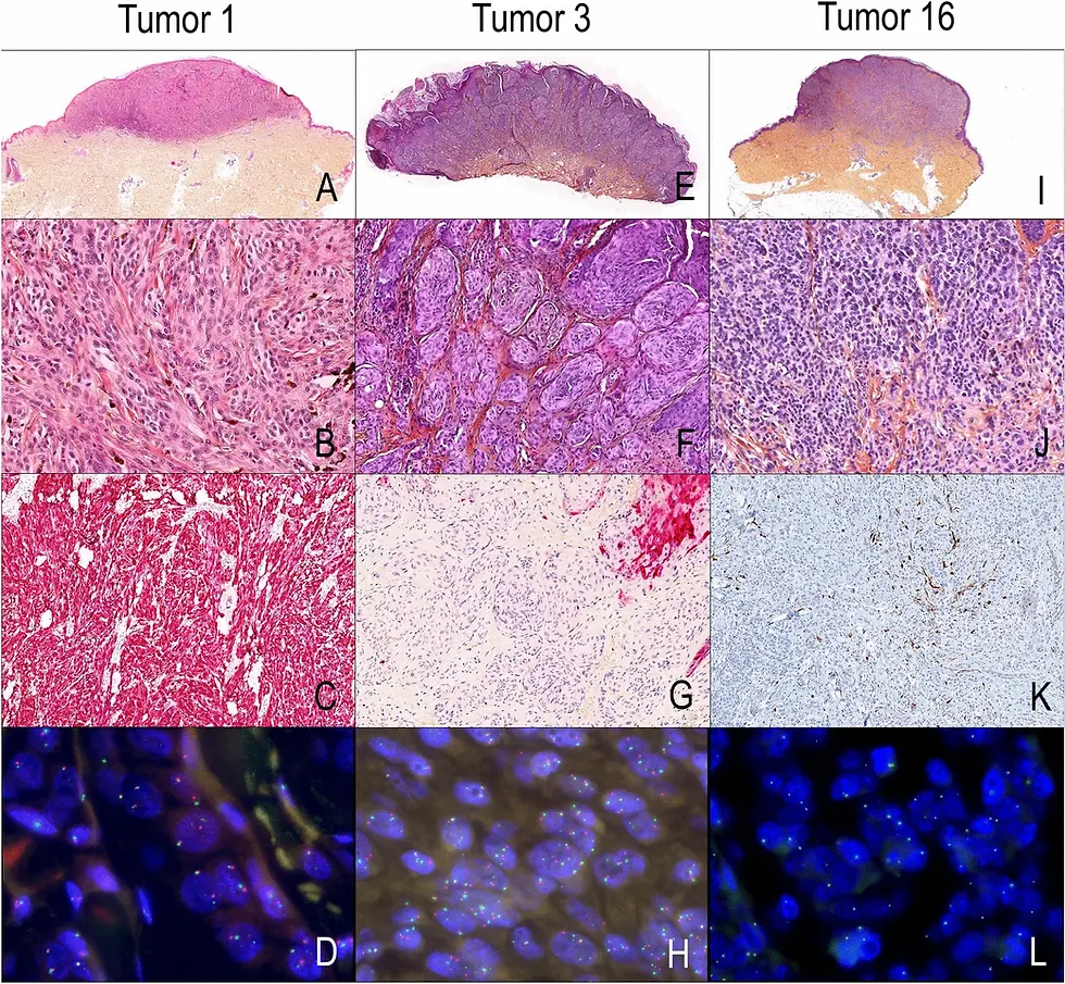
Researchers retrospectively analysed 206 atypical melanocytic tumours submitted for expert second opinions. They evaluated two key laboratory techniques:
p16 IHC, detecting the protein encoded by the tumour suppressor CDKN2A — often lost in melanomas.
9p21 fluorescence in situ hybridization (FISH), which identifies homozygous deletions at the 9p21 locus, a common genomic alteration in malignant melanocytic lesions.
By comparing initial histopathological diagnoses with expert reviews that included p16 IHC and 9p21 FISH results, the authors developed and tested a simple diagnostic algorithm for routine clinical practice.
Key findings
The study found that:
Negative p16 IHC strongly correlated with malignant diagnoses: 90% of malignant cases showed p16 loss, while only 11% of benign tumours did.
Homozygous 9p21 deletion was highly predictive of malignancy, showing strong agreement with expert histopathological diagnosis when present.
Combining IHC and 9p21 FISH enhanced diagnostic confidence, helped upgrade uncertain diagnoses, and clarified tumour classification in challenging cases.
Importantly, the authors observed that heterozygous 9p21 deletion lacked diagnostic value, and that p16 loss alone could suggest malignancy but did not always confirm it unless accompanied by homozygous deletion.
Why these results matter to histology researchers
Although this is a clinical pathology study, there are several key lessons for histology research workflows — especially for those using IHC and other tissue biomarkers:
1. High-quality IHC remains central
The value of p16 IHC in this study underlines the power of carefully validated immunohistochemical staining. Reliable antigen detection and interpretation can dramatically impact diagnostic outcomes, especially in ambiguous or borderline lesions — highlighting why robust IHC procedures are essential for research as well as clinical work.
2. Integration with molecular tools enriches interpretation
While histological morphology and IHC can carry much of the diagnostic weight, integrating molecular genetics (such as FISH) provided added confidence in the study. For research, this suggests that multimodal tissue analysis — combining IHC with other molecular insights — can improve interpretation of complex biological phenomena.
3. Algorithms based on biomarkers improve reproducibility
The proposed algorithm starts with p16 IHC and adds 9p21 FISH only when p16 is negative. A structured approach like this helps standardise interpretations and reduces inter-observer variability — a principle that’s equally useful in research settings where reproducibility is crucial.
What this means for cancer and tissue research
This study reinforces the importance of marker selection and interpretation in histology. Even outside diagnostics, research frequently faces similar challenges, such as:
Differentiating tumour subtypes
Validating tumour suppressor involvement
Establishing correlations between protein expression and genetic alterations
In such contexts, IHC remains one of the most accessible and informative tools for tissue research. Combining IHC with additional molecular assays — whether FISH, RNA probes, or next-generation sequencing — broadens the analytical context and depth.
How LabNexus supports research applications of histology
At LabNexus, we provide a range of histology services that help researchers explore tissue biology with precision and clarity. Our offerings include:
High-quality immunohistochemistry (IHC) for research markers, including p16, Ki-67, and many others
Single-plex or multi-colour chromogenic IHC, including DAB and coloured chromogen options
Multiplex IHC panels that allow multiple markers on the same tissue section
Tissue processing, paraffin embedding, and sectioning
High-resolution slide scanning for digital and quantitative analysis
Our state-of-the-art platforms and trained histotechnologists ensure consistent, reproducible staining results — enabling researchers to generate robust data that supports publication, biomarker discovery, or translational studies.
Note: LabNexus provides research-only histology services, and does not perform diagnostic testing.
Conclusion
The Scientific Reports study highlights how combining p16 IHC and 9p21 FISH improves diagnostic accuracy in challenging melanocytic tumours. For research scientists, it reinforces key lessons:
The importance of validated IHC protocols
How combining markers enhances confidence in interpretation
The value of structured biomarker algorithms
Histological techniques continue to play a central role — not just in diagnostics, but in deepening our understanding of tissue biology and disease mechanisms.
References
Vergara, R., Laharanne, E., de la Fouchardière, A., et al. (2025). Improving diagnostic accuracy in atypical melanocytic tumors using p16 immunohistochemistry and 9p21 fluorescence in situ hybridization: analysis of 206 second opinion cases. Scientific Reports, 15, 11425.


Comments