.png)
How Do Histology Stains Help Research & Patient Diagnosis?
- Eghosa Arovo

- Jan 5
- 3 min read
Curious how tissue stains do more than just add colour? Discover how they unlock insights in research and play a key role in diagnosis.

Introduction
In histology, stains are more than just colour—they’re tools for discovery. By highlighting specific structures, stains enable researchers and clinicians to explore the fine details of tissues and cells. In research, staining techniques support studies in cancer biology, tissue development, and regenerative medicine. In clinical settings, they help pathologists diagnose and classify disease.
In this blog, we’ll explore how common stains are used in research and diagnosis, with a focus on how researchers can leverage these techniques to generate deeper insights from their tissue samples.
How Stains Support Research
1. Understanding Tissue Architecture
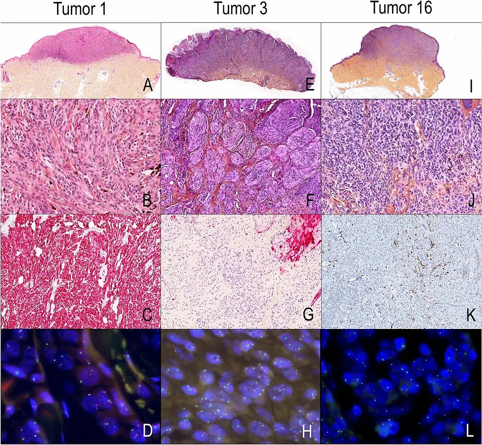
Routine stains such as Haematoxylin and Eosin (H&E) remain the foundation of tissue analysis. Haematoxylin stains cell nuclei blue-purple, while eosin colours the cytoplasm and extracellular matrix in shades of pink. This combination allows researchers to assess overall tissue structure, identify tumour margins, and evaluate abnormal tissue growth.
2. Targeting Specific Tissue Components
Special stains are used to highlight particular elements within tissue:
Sirius Red binds to collagen, revealing areas of fibrosis or extracellular matrix changes.
Perls’ Prussian Blue detects ferric iron deposits, useful in studies of iron metabolism or haemorrhage.
Reticulin and EVG (Elastic Van Gieson) stains show structural fibres like reticulin and elastin, aiding in vascular or connective tissue analysis.
DPAS (Diastase-PAS) differentiates glycogen from other carbohydrates.
Masson’s Trichrome distinguishes muscle, collagen, and cellular components, often used in fibrosis and invasion studies.
These stains allow researchers to go beyond general structure and study the biochemical makeup of tissues in cancer models, fibrosis studies, and organoid development.
3. Investigating Cell Phenotypes with IHC
Immunohistochemistry (IHC) uses targeted antibodies to detect specific proteins within tissue. This technique is crucial in research areas like tumour immunology, where it helps identify immune cell types (e.g. CD3, CD8), proliferation markers (e.g. Ki-67), or oncogene expression (e.g. HER2).
For example, researchers studying the tumour microenvironment can use IHC to map immune cell infiltration, compare response to treatment, or assess changes in stromal signalling.
4. Turning Colour into Data with Digital Imaging
With digital slide scanning, stains are no longer only visual—they become data. Image analysis software can quantify staining intensity, measure fibrosis area, or count specific cell populations. This quantitative approach improves reproducibility and enables statistical comparisons across experimental groups.
The Role of Stains in Diagnosis
While LabNexus focuses exclusively on research, it’s worth understanding how similar staining methods are used in diagnostic labs. Pathologists use H&E and special stains to classify tumours, detect infections, or identify structural abnormalities. IHC is routinely used to determine tumour subtypes or to assess biomarkers that guide treatment decisions.
Although our work does not involve diagnosis, we apply the same level of precision and quality that diagnostic labs use—helping researchers produce translational data that can model clinical outcomes.
Conclusion
Histological staining remains a cornerstone of tissue-based research. Whether you're visualising tumour margins, mapping fibrosis, or identifying specific cell populations, stains provide both clarity and context. For research students and investigators in cancer biology and tissue studies, understanding the strengths of each stain can significantly enhance the depth and quality of your findings.
If you're planning a project that involves histology, LabNexus offers expert research-only services—including processing, embedding, sectioning, H&E, special stains, IHC, and slide scanning.
Book a free consultation today to discuss how we can support your next research milestone.
References
Bancroft, J.D. & Gamble, M. (2020). Theory and Practice of Histological Techniques (8th ed.). Elsevier.
Puchtler, H., Waldrop, F.S., & Valentine, L.S. (1965). A study of the specificity of the reticulin stain. Journal of Histochemistry & Cytochemistry, 13(4), 275–285.
Junqueira, L.C., Bignolas, G., & Brentani, R.R. (1979). Picrosirius staining plus polarization microscopy for collagen visualization. Histochem J, 11(4), 447–455.
Elgsaeter, A. & Higman, A. (1977). Improved detection of ferric iron using Perls’ Prussian Blue method. Journal of Histochemistry & Cytochemistry, 25(4), 367–369.
Pearse, A.G.E. (1985). Histochemistry: Theoretical and Applied (4th ed.). Churchill Livingstone.


Comments